Dansk  Webmaster  Search   


AS, General aspects
AS, Sacroiliitis - radiography
AS, Sacroiliitis - MRI
AS, Spine - radiography
AS, Spine - MRI
AS, Spinal fractures
AS, Other joints
Other forms of SpA
Psoriatic arthritis
Reactive arthritis
Enteropathic arthritis
Undifferentiated SpA
PAO / SAPHO / CNO
Juvenile spondyloarthritis
General aspects

All the different forms of axSpA seen in adults can occur in children/adolescents, but with a predominance of undifferentiated axSpA, doi:10.3899/jrheum.180168. However, if the skeleton has not reached maturity when involved by inflammation there are usually concomitant growth-related changes which have to be taken into account as shown beneath.

 
SpA in childhood

MRI of the SIJ in a 13-year-old boy with inflammatory low back pain and normal spinal MRI, semi-coronal T1 and T1FS, semi-axial STIR and straight coronal STIR image showing subchondral edema at the right SIJ (thick arrows) in addition to growth-related features in the form of nuclei in the joint spaces (open arrows) and a persistent disk between the first and second sacral segment (long slim arrow).

Four years later at the age of 17 years, MRI shows bilateral subchondral edema, most pronounced on the left side (thick arrows). There is concomitant edema at the persistent intervertebral space between S1 and S2 (long slim arrow).

Concomitant T1 and T1FS images show manifest structural changes in the form of erosions, especially of the iliac joint facets (thick arrows), with dispersed fat deposition, but also irregularity with adjacent fat deposition at the persistent intervertebral space between S1 and S2 (long slim arrows). Besides, there is persistence of intraarticular not fused nuclei (open arrows).

 
axSpA changes like in adulthood

MRI in a 16-year-old girl presenting with non-radiographic active sacroiliitis, coronal T1, T1FS and axial STIR image showing pronounced subchondral edema at the right SIJ and slight edema at the left SIJ (arrows), but no structural changes.

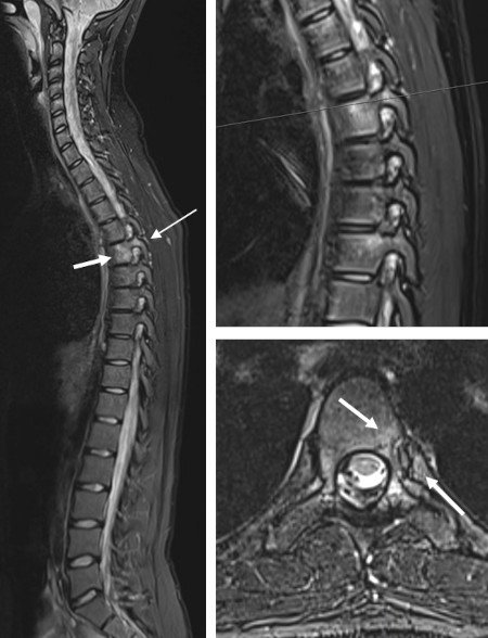
Concomitant spinal MRI, sagittal STIR of the whole spine with enlargement of the thoracic region to the right with insertion of the scan level for the supplementary axial STIR image shown below. There is edema at two costovertebral joints (arrows), at the upper joint extending to the apophyseal joint, changes conforming to the early axSpA changes seen in adulthood.

 
Visitors since May 1st., 2008:  1768424