Psoriatic arthritis is a chronic inflammatory rheumatic disease affecting patients with the dermal disease psoriasis.
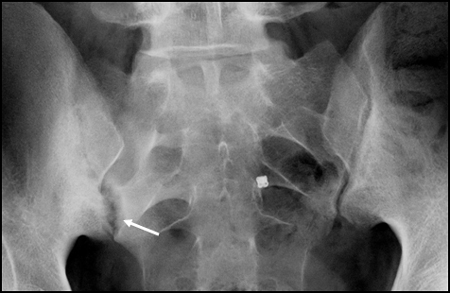
Up to 20-25% of patients with psoriasis have psoriatic arthritis (PsA) with the highest frequencies occurring in patients with moderate to severe psoriasis, doi:10.1016/J.JAAD.2018.06.027. Involvement of the axial skeleton (sacroiliac joints and spine) is a relatively frequent manifestation, mostly together with peripheral arthritis, enthesitis and/or dactylitis.
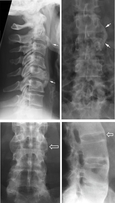
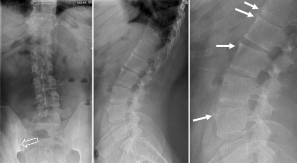
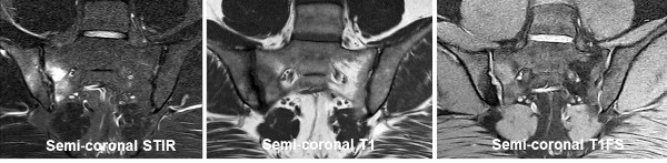
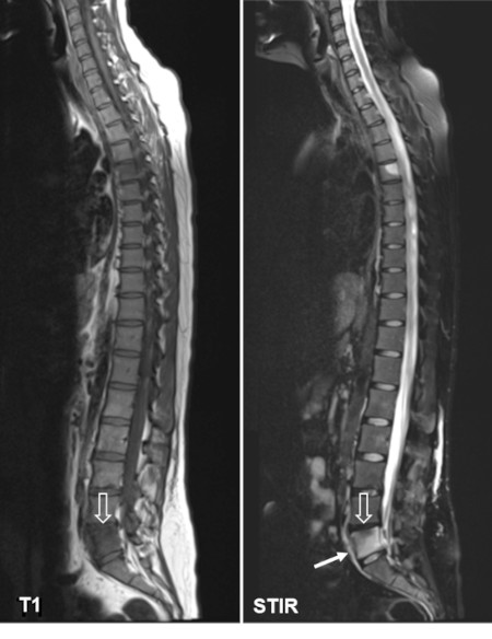
Some features typical for axial involvement in PsA are compared with axial SpA without psoriasis somewhat different such as lower prevalence of inflammatory back pain and HLA-B27, and the occurrence of isolated involvement of the spine without concomitant SIJ changes in up to 30% of the patients with axial PsA, doi: 10.1136/ANNRHEUMDIS-2016-209853. The morphology of the syndesmophytes is often different from ypical syndesmophytes in AS, being asymmetrical, non-marginal and more voluminous/chunky doi: 10.31138/MJR.33.1.142. However, a clear distinction between axial PsA and axSpA without psoriasis is not always possible due to a natural overlap between these conditions.
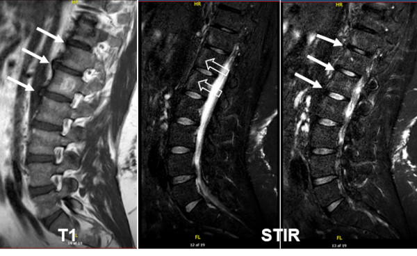
According to the ASAS criteria, patients with axial PsA can be classified as patients with axSpA if fulfilling the ASAS criteria, presenting chronic back pain with onset prior to the age of 45 years plus presence of sacroiliitis by MRI or radiography and one additional SpA feature that can be psoriasis, or alternatively having HLA-B27 plus 2 additional SpA features. However, PsA patients with axial involvement may have characteristics that do not allow an axSpA classification such as late onset of back pain, involvement of the spine without SIJ changes and lack of HLA-B27, doi:10.1136/ARD.2008.104018.
There are no widely accepted criteria for axial involvement in PsA. Therefore, clinical and imaging manifestations indicative of axial PsA are currently the subject of a large international study including imaging encompassing radiography and MRI of the SIJ and spine, doi:10.1177/1759720X211057975.
Because symptoms and signs of axial involvement often occur at older ages than AS, the imaging features can be mixed with degenerative and/or load-related changes. |