Dansk  Webmaster  Search   


AS, General aspects
AS, Sacroiliitis - radiography
AS, Sacroiliitis - MRI
AS, Spine - radiography
AS, Spine - MRI
AS, Spinal fractures
AS, Other joints
Other forms of SpA
Psoriatic arthritis
Reactive arthritis
Enteropathic arthritis
Undifferentiated SpA
PAO / SAPHO / CNO
Juvenile spondyloarthritis
General aspects

Reactive arthritis is usually a transient acute arthritis appearing a few weeks after an infection somewhere in the body, mostly located in the urinary or the gastrointestinal tracts. It usually involves large joints, such as knees and ankles as well as the feet, but there may also be inflammatory changes located to the SIJ and/or spine in the acute stage (as shows below) as well as extra-musculoskeletal manifestation such as conjunctivitis, keratoderma blenorrhagicum, urethritis or erythema nodosum.

The disease usually vanishes within a year, but may recur and up to 30% develop a chronic condition. This is especially the case in patients with the tissue type HLA-B27. Patients with a chronic course often have involvement of the sacroiliac and/or spinal joints conforming to axSpA.

There is little evidenced knowledge about the imaging differences or similarities of the changes in reactive arthritis compared to AS. Although unilateral sacroiliitis and non-marginal syndesmophytes has been reported in reactive arthritis the condition can develop into AS like changes, especially in HLA-27 positive patients (doi:10.1136/ARD.57.3.135).

It seems that the SpA-/B27-related reactive arthritis is decreasing in incidence which can be due to less food related diseases, cleaner water, quicker treatment of infections, such as sexually transmitted infections, or mutation of common infectious agents of reactive arthritis (Salmonella, Shigella, chlamydia etc.) with less changes of producing an immunological response. Other post-infectious arthritides elicited by a wide variety of organisms have been described, but only few of them seems related to axSpA, doi:10.1007/S10067-018-4022-5; doi:10.1007/S11926-021-01018-6.

 
Sacroiliac joints

The active changes may be asymmetrical and are usually more limited than seen in AS. In uncomplicated cases the subchondral edema is transient and can vanish without the development of structural changes as shown below. However, persistent inflammation will often result in concomitant structural changes. A special feature which can occur in reactive sacroiliitis is concomitant edema in the soft tissue which can simulate infectious changes necessitating postcontrast sequences, as shown beneath. In case of transition to a chronic stage, the SIJ changes will often have features consistent with those of AS.

 
Sacroiliac joint - acute changes

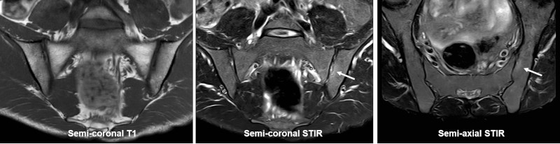
MRI scan of the SIJ in a young man who, after a chlamydia infection 2 months previously, develops knee joint arthritis and left-sided buttock pain; semi-coronal T1 and STIR, and semi-axial STIR image show slight subchondral edema in the ileum at the left SIJ, as a sign of active inflammatory changes. There are no visible structural changes and the symptoms of sacroiliitis disappeared within 2 months.

 
Sacroiliac joint – mixture of active and structural changes

MRI, in a patient with a urogenital Clamydia infection one year previously and persistent symptoms of sacroiliitis. Semi-coronal T1, T1FS and STIR in addition to semi-axial STIR showing pronounced subchondral edema at the left sacroiliac joint (arrows) in addition to edema in the soft tissue anteriorly (open arrow). There is also erosive changes with concomitant subchondral fat deposition (asterisk).

 
Spine

Spinal changes are relatively frequent in chronic disease. They may vary in appearance depending on disease severity and activity. There is sometimes more extensive new bone formation than seen in AS with concomitant osseous changes corresponding to the attachment of non-marginal syndesmophytes as shown below, but the ossifications may be difficult to detect by sagittal MR sequences. However, chronic reactive arthritis may transform to an AS-like disease with imaging features characteristic of AS. This is especially seen in patients with the tissue type HLA B27.

Radiographs, frontal and lateral view, showing structural changes in the form of new bone formation around the discs which are more voluminous than generally seen in AS (arrows).

MRI, sagittal T1 and STIR image, showing hypertrophic ligamentous ossification at the intervertebral space Th11-Th12 and possible ossification in the lumbar region (arrows). There is concomitant fat deposition anteriorly in the Th11 vertebral body (open arrow on the T1 image) with a brim of edema posteriorly and also a small edematous area anteriorly in the Th12 vertebral body (open arrows on the STIR image).

 
Visitors since May 1st., 2008:  1768448