Dansk  Webmaster  Search   


AS, General aspects
AS, Sacroiliitis - radiography
AS, Sacroiliitis - MRI
AS, Spine - radiography
AS, Spine - MRI
AS, Spinal fractures
AS, Other joints
Other forms of SpA
Psoriatic arthritis
Reactive arthritis
Enteropathic arthritis
Undifferentiated SpA
PAO / SAPHO / CNO
Juvenile spondyloarthritis
General aspects of spinal fractures

The spine gradually fuses in patients with ankylosing spondylitis (AS) through ligamentous ossification, syndesmophytes and ankylosis of apophyseal joints resulting in a rigid hyperkyphotic deformity. Biomechanically the fused bones are like a long bone and the rigidity makes the spine incapable of appropriately dissipating the energy of a traumatic event. The altered spinal biomechanics combined with a frequent occurrence of brittle osteoporotic bones increase susceptibility to vertebral column fractures, even after minor often trivial trauma.

 
Fracture types

In AS and other axSpA forms two different types of fractures may occur: compression fractures as part of osteoporosis and transverse fractures.

 
Osteoporotic compression fractures

May occur in all stages of the disease and contribute to the thoracic kyphosis often seen in AS. These fractures are usually stable and rarely cause spinal cord injury, though impingement of the cord may occur.

MRI of the thoracic and cervical spine (T1 and STIR image) in an AS patient with thoracic kyphosis complaining of progressive pain in the lower thoracic region without preceding trauma. There is pronounced osteoporotic collapse of Th12 (arrows) with osseous impingement on the spinal cord (open arrow).

 
Transverse fractures

Transverse fractures mainly occur in ankylosed spines, seen in late stages of AS. They are caused by trauma, but occasionally patients do not remember having had a trauma as it can be minimal and result from a fall from standing or seating position.

Transverse fractures are mostly located to the lower cervical and upper thoracic spine. They pass through ankylosed intervertebral spaces or vertebral bodies and will not always respect normal anatomical cleavages. They may cross the spine horizontally or obliquely through several vertebral bodies. The posterior structures are usually involved, including the apophyseal joint region and ossified ligaments making the fracture site unstable. Thus, fractures of the stiff spine resemble fractures of tubular bones with a potential for dislocation. In the spine, this instability carries a risk for serious spinal cord or nerve injuries.

It is important to bear in mind that in an ankylosed spine with a fracture movements will occur at the fracture site. This can cause serious adverse events, particularly in the cervical region, if the fracture is attempted stabilized in a cervical collar which does not fit. The spinal cord can thereby be compressed, resulting in neurological lesions following the movement as illustrated below.

Fracture of C7 - before applying a cervical collar - with only slight dislocation of the fracture. The figure is shown with kind permission from Acta Orthop Belg 2010;76:413-5.

Fracture of the C7 – after applying a cervical collar. The fracture has been torn apart and there is angulation corresponding to the spinal canal potentially causing damage to the spinal cord. The figure is shown with kind permission from Acta Orthop Belg 2010;76:413-5.

 
Patient recommendation

The Danish (and other national) Ankylosing Spondylitis Society and the Ankylosing Spondylitis International Federation (ASIF) recommend that patients carry information cards about their illness, including a photo showing the normal posture of the person. This must be considered by paramedics, emergency room physicians, radiographers etc. in cases of suspected fracture to ensure adequate spinal stabilization.

Information card recommended by The Danish Ankylosing Spondylitis Society.

Medical recommendations

When possible, it is important to diagnose fractures before they are dislocated and potentially giving rise to neurological deficits. Hence the following guidelines.


The possibility of spinal fracture must always be considered when an AS patient reports:

  • A direct or indirect spinal trauma, even minor trauma.
  • Newly onset of mechanical back pain that is triggered or worsened by activity and completely or partially relieved by rest.
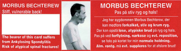
  • Changes in posture; if necessary, check photo of the patient on the information card.
  • Occurrence of new neurological symptoms (even weak). These can occur immediately after a trauma or be delayed for hours or days. 


When substantive suspicion of fracture is present, the following is important:

  • Manage the patient as having an unstable fracture until the diagnosis is excluded.
  • Avoid the use of rigid cervical collar, if it does not immediately fit the patient's shape.
  • Do not attempt to correct a kyphotic spine.
  • When positioning and moving the patient, support the entire spine in the position present before the trauma.


Diagnostic Imaging:

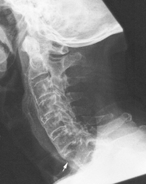
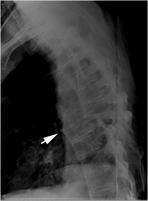
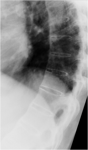
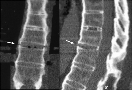
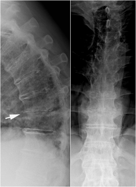
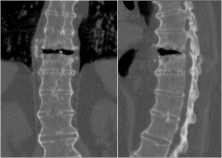
Conventional radiography in at least two planes is often the initial examination. It can be used to diagnose osteoporotic compression fractures and show obvious transverse fractures.


Obvious fracture through a discus and the posterior structures with dislocation of the lower part of the spine anteriorly to the upper part of the spine above the fracture site (arrow).


Altered anatomy with poorly defined discs, osteoporosis, calcified ligaments etc. often complicate interpretation of conventional radiographic images. A non-dislocated fracture can be difficult to diagnose with certainty by conventional radiography. There may only be a small osseous break in a syndesmophyte although there is a potentially unstable fracture, which may dislocate and cause neurological deficits.

AS patient with pronounced pain after a tennis game. Radiography does not confirm the occurrence of a fracture. There is no visible malalignment or osseous breaks.

Supplementary CT, coronal and sagittal 2D reconstructions, shows fracture corresponding to syndesmophytes with increased height of the intervertebral space aneriorly, signs compatible with a horizontal fracture.


Conventional radiography can thus only contribute diagnostically if there are positive findings such as obvious fracture or e.g., visible misalignment between the vertebrae. A simple fracture of a spinosus process also points with high probability to the presence of a transverse fracture, doi:10.1007/s00256-008-0484-x


Standing radiography of the thoracic spine in a 64-year-old man with AS and persistent mechanical back pain during 3-4 weeks. There is slight displacement between Th9 and Th10 anteriorly (arrow), and the intervertebral space has reduced height both on the lateral and frontal view, all suggesting fracture.

CT performed in supine position, coronal and sagittal reconstructions clearly show the fracture through the intervertebral space and the posterior structures. In this position the intervertebral space opens anteriorly with concomitant reduced space in the spinal canal.


It is important to be aware that conventional radiography cannot exclude the presence of spinal fracture. Supplementary computer tomography (CT) and/or magnetic resonance imaging (MRI) may therefore be necessary. Besides, most fractures are cervical or at the cervico-thoracic transition where fractures can be difficult to detect by radiography due to superimposed shoulders. Also the occasional occurrence of pronounced kyphosis can impede an optimal frontal view. In this region CT may therefore be the preferred initial examination.

 
Computer tomography
Is often necessary to confirm the diagnosis of transverse fractures and/or delineate the osseous structure before surgical stabilization. CT is superior to conventional radiography in the diagnosis of fractures by giving the possibility of multiplanar reconstructions in relevant planes. Be aware that there may be more than one fracture, especially in patients with obvious trauma.

AS patient after a fall from a bicycle. Primary CT shows two transverse fractures in the cervical region (arrows).

 
Magnetic resonance imaging

MRI is always indicated in patients with neurologic deficits. It can visualize spinal cord injury and hemorrhages, most importantly epidural hemorrhage, often necessitating emergency surgery doi:10.1007/s00256-008-0484-x; doi:10.2214/AJR.08.1616.

Lateral radiograph and MRI (sagittal T1 and T2 image) of the cervical spine in an AS patient with neurological deficits (paresis) after a trauma. There is a transverse cervical fracture through the intervertebral space and the vertebral body of C5 with angulation at the fracture site (white arrows). Besides the fracture, there is hemorrhage in the spinal cord corresponding to the angulation area (black arrow). Part of the spinal cord damage may be caused by inappropriate stabilization in a position “not normal" for the patient; please see the first patient illustrated with transverse fracture.

Visitors since May 1st., 2008:  1758431