Dansk  Webmaster  Search   


AS, General aspects
AS, Sacroiliitis - radiography
AS, Sacroiliitis - MRI
AS, Spine - radiography
AS, Spine - MRI
AS, Spinal fractures
AS, Other joints
Other forms of SpA
Psoriatic arthritis
Reactive arthritis
Enteropathic arthritis
Undifferentiated SpA
PAO / SAPHO / CNO
Juvenile spondyloarthritis
Diagnosis of spinal AS changes by radiography

Conventional radiography is a well-established method for detecting structural spinal changes such as new bone formation between the vertebral bodies and vertebral fusions. In manifest disease radiography often gives a good overview of structural changes.


For the diagnosis of spinal changes lateral as well as frontal radiographs are needed. However, despite using two planes the thoracic spine can be difficult to evaluate on radiographs due to overlaying structures such as lung tissue. Supplementary MRI may be needed in early stages and for monitoring the disease during therapy. MRI can display signs of activity which cannot be obtained by radiography.


If fracture is suspected, but cannot be diagnosed or excluded by radiography supplementary CT may be necessary and also MRI in the case of spinal cord or nerve lesion.


The radiographic AS changes vary with the disease duration and severity as illustrated below.

 
Slight early changes

Anterior and lateral radiographs of the lumbar spine and thoraco-lumbar junction showing slight vertebral squaring at the thoracolumbar junction (arrows) with slight osseous condensation anteriorly in the vertebral bodies. The anterior view shod manifest bilateral sacroiliitis changes.

 
Manifest early changes

Frontal and lateral radiographs of the lumbar spine showing a bridging syndesmophyte (black arrow), sclerosis at the upper corner of L1 (white arrow) and L2 and vertebral squaring at the thoracolumbar junction. Besides, a slight osseous deposition at the anterior surface of the first lumbar vertebral body (L1)(arrowhead), which can contribute to the formation of vertebral squaring. 

 
Manifest changes

Frontal and lateral radiographs of the lumbar spine showing bridging syndesmophytes (arrows) between several vertebral bodies (arrows).

 
Advanced changes

Frontal and lateral radiographs of the lumbar spine showing fusion of several vertebral bodies (arrow) in addition to fusion of the apophyseal joints and ossification of the interspinous ligament posteriorly.

 
Grading methods


Assessment of the severity of radiographic spinal abnormalities (grading) in AS is usually performed in consistence with the internationally recommended SASS or modified SASS (mSASSS) method,

Doi:10.1093/rheumatology/key128.


SASSS = Stoke Ankylosing Spondylitis Spine Score:


Anterior and posterior changes in the lumbar spine (Th. 12 - S1) at lateral view.

  • Grade 0: Normal findings.
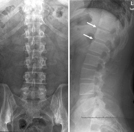
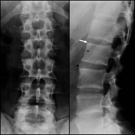
  • Grade 1: Erosion and/or sclerosis and/or squaring.
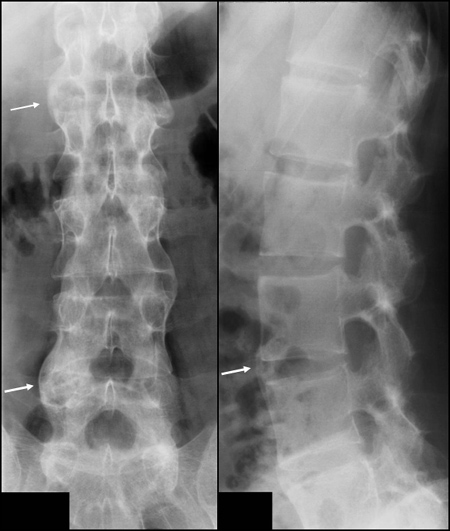
  • Grade 2: Syndesmophytes (non-bridging).
  • Grade 3: Ankylosis (total bone bridging between upper and lower vertebral edges.


Modified SASSS (mSASSS)

Only anterior changes in both the cervical and lumbar spine at lateral view. Is recommended by OMERACT.

 
Visitors since May 1st., 2008:  1767424