Dansk  Webmaster  Search   


AS, General aspects
AS, Sacroiliitis - radiography
AS, Sacroiliitis - MRI
AS, Spine - radiography
AS, Spine - MRI
AS, Spinal fractures
AS, Other joints
Other forms of SpA
Psoriatic arthritis
Reactive arthritis
Enteropathic arthritis
Undifferentiated SpA
PAO / SAPHO / CNO
Juvenile spondyloarthritis
Typical MR changes in ankylosing spondylitis

Typical MR changes in ankylosing spondylitis depend on the disease stage.


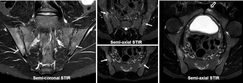
Early stages: Signs of disease activity in the form of edema and/or contrast enhancement in the subchondral bone. The changes are often bilateral, but may be unilateral and vary from side to side over time. To establish an AS diagnosis there need to be accompanying structural changes in the form of erosion, joint space alteration and/or fatty deposition in the bone marrow to fulfill the radiographic New York criteria.


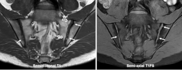
Middle stages: Is often dominated by structural changes (erosion, joint space alteration in the form of narrowing or widening and fat deposition in the bone marrow). During active stages the structural changes are mixed with signs of activity (edema and/or contrast enhancement), which can vary from side to side.


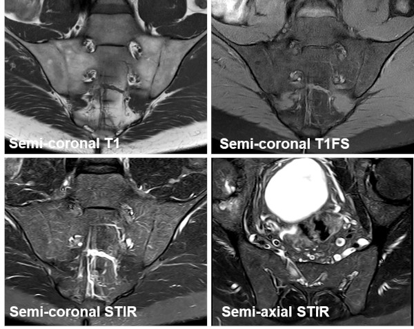
Late stages: Increasing joint destruction with partial or complete fusion (ankylosis) of the joints, which can appear as "ghost joints" (ankylosis with visible joint contours).

 
Early ankylosing spondylitis - activity

Semi-coronal and semi-axial STIR images in a 22-year-old, HLA-B27 positive, man with radiographic sacroiliitis, right SIJ grade 2, left SIJ grade 3. There are manifest signs of activity in the form of subchondral edema (arrows) at both SIJ. The semi-axial STIR image to the right with a large field of view, including the symphysis, reveal concomitant active enthesitis changes at the left pubic bone (open arrow).

Early ankylosing spondylitis - structural changes

Concomitant semi-coronal T1 and T1FS image show obvious erosion of both joints (arrows) and also a brim of subchondral fat deposition on the left side (open arrows).

 
Manifest ankylosing spondylitis – mixture of active and structural changes

Manifest AS in a 54-year-old, HLA-B27 positive, man with symptoms since the age of 24 years; semi-coronal T1, T1FS and STIR in addition to semi-axial STIR with a large field of view including the symphysis. There are signs of disease activity in the form of subchondral edema, especially on the left side (white arrows), but also structural changes in the form of erosions and joint space narrowing with ligamentous ankylosis proximally (black arrows) and pronounced subchondral fat deposition, especially in the sacrum (open arrows). There are concomitant small subchondral edema areas in the left pubic bone (long thin arrow).

Late stage - ankylosis

Late stage semi-coronal T1-weighted, T1FS and STIR in addition to a semi-axial STIR image show obliteration of the SIJs with fat signal across the joint spaces, which are discernable on the T1-weighted sequence (ghost joints). There is no evidence of activity on the STIR image.

 
MRI grading methods

Grading of inflammatory changes at the SIJs is used to monitor the disease, especially in scientific studies. It is today usually performed in accordance with the SPARCC or Berlin scoring methods described in the section about MRI (Grading sacroiliitis by MRI).

 
Visitors since May 1st., 2008:  1767951