Dansk  Webmaster  Search   


AS, General aspects
AS, Sacroiliitis - radiography
AS, Sacroiliitis - MRI
AS, Spine - radiography
AS, Spine - MRI
AS, Spinal fractures
AS, Other joints
Other forms of SpA
Psoriatic arthritis
Reactive arthritis
Enteropathic arthritis
Undifferentiated SpA
PAO / SAPHO / CNO
Juvenile spondyloarthritis
Diagnosis of changes in other joints

All other joints can be involved by ankylosing spondylitis, but joints close to the trunk are most frequently involved, these are the hip, shoulder and joints in the anterior chest wall. The changes are characterized by joint destruction with erosion accompanied by new bone formation at attachments of tendons and/or joint capsules (enthesopathy). Manifest changes in the shoulder and hip joints are often well visualised by conventional radiography. However, other imaging methods may be necessary.

CT and/or MRI has a central role in detecting anterior chest wall changes which can be difficult to visualise by radiography.

In the case of normal pelvic radiography MRI may provide information about active inflammation as well as erosion and enthesopathy.

Ultrasonography is useful for detecting joint fluid accumulation and edema in the soft tissue, but cannot visualize osseous changes.

Scintigraphy and/or PET may also be used for detecting active inflammation, especially if it is necessary to get an overview of the entire body. However, there examinations are associated with a certain amount of radiation and will usually not like MRI visualize disease abnormalities in detail. They are therefore in frequently used today.

 
Radiographic enthesopathy
Enthesopathy, frontal pelvic radiograph in a patient with AS showing pronounced new bone formation at the ischial bones (arrows) in addition to ankylosis of the SIJs and the pubic symphysis. There are also changes in the left hip consistent with osseous necrosis (open arrow).
 
Active enthesopathy - MRI

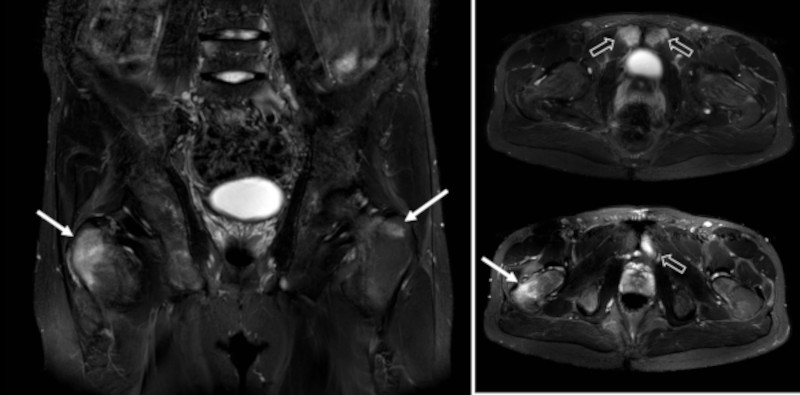
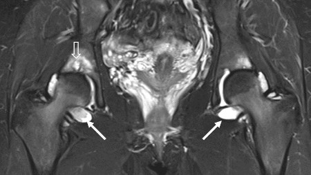
MRI, coronal and axial STIR images in a patient with bilateral sacroiliitis showing active enthesopathy changes at the greater trochanter on both sides (arrows), most pronounced on the right side. There is also osseous edema at the pubic symphysis (open arrows).

 
Hip synovitis - MRI

Coronal STIR of the hips in a patient with bilateral sacroiliitis showing pronounced synovitis in both hips (arrows) and also erosive changes (open arrow).

 
Anterior chest wall involvement

CT of the sternum and surrounding joints. 2D (left image) and 3D reconstructions show fusion of the joint between the upper and lower parts of the sternum. In addition, erosion corresponding to the joints between the sternum and the clavicle (sterno-clavicular joints) and exuberant mineralization of the costal cartilages, especially the two upper cartilages.

 
Visitors since May 1st., 2008:  1767821