Dansk  Webmaster  Search   


AS, General aspects
AS, Sacroiliitis - radiography
AS, Sacroiliitis - MRI
AS, Spine - radiography
AS, Spine - MRI
AS, Spinal fractures
AS, Other joints
Other forms of SpA
Psoriatic arthritis
Reactive arthritis
Enteropathic arthritis
Undifferentiated SpA
PAO / SAPHO / CNO
Juvenile spondyloarthritis
Diagnosis of spinal AS changes by MRI

Magnetic resonance imaging (MRI) can often give valuable information in the early stage of spinal involvement providing evidence of active inflammation, which cannot be seen in other ways. Structural changes can also be detected and MRI can be useful for monitoring the disease including the effect of therapy.

 
Typical AS changes

Typical MR changes in ankylosing spondylitis depend on the disease stage as briefly stated and illustrated beneath, but the changes can vary considerably and may be modified by treatments.


Early stage: Signs of disease activity in the form of bone marrow edema (BME) typically located to the vertebral corners and/or at the joints between thoracic vertebral bodies and the ribs (costovertebral joints). There may be active inflammation in other locations, typical for AS inflammation such as apophyseal joints, costotransverse joints and interspinous ligaments although usually seen later in the disease.


Middle stage: Often a mixture of signs indicating activity as in early stages and structural/chronic changes in the form of fat deposition in the bone marrow corresponding to previous BME areas and squaring of vertebrae due to destruction/erosion of the vertebral corners. Syndesmophytes are only visible if edematous or containing fatty tissue.


Late stage: More pronounced, structural changes with bridging syndesmophytes or complete fusion of the vertebral bodies. During active phases there is often concomitant signs of disease activity.

 
Typical sites of AS changes in the spine

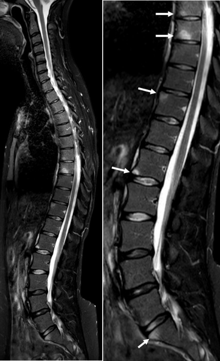
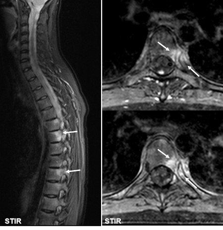
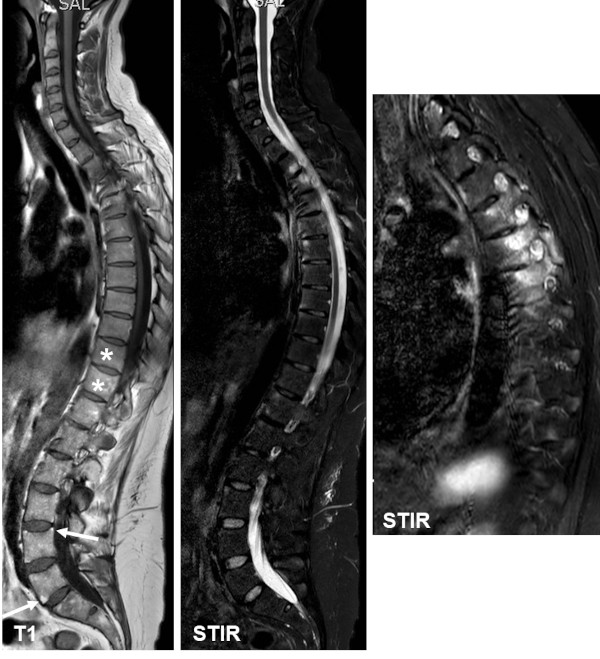
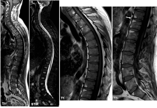
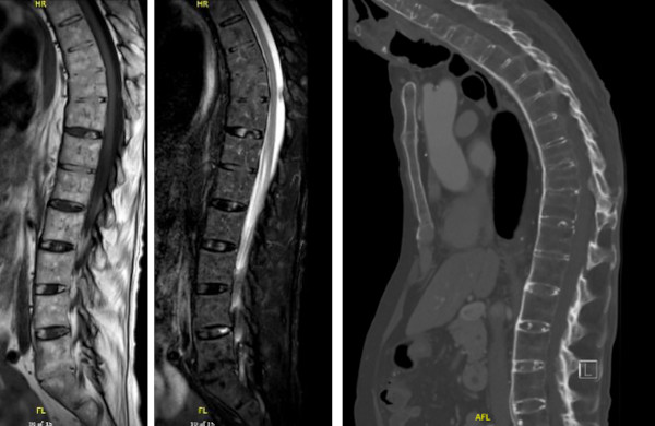
Irrespective the stage of the disease, there are typical locations for active inflammatory and structural spinal changes in AS by MRI encompassing:

·      BME/osteitis and/or fat deposition at vertebral corners, located to one or more of the four vertebral corners on sagittal images

·      Syndesmophytes new bone formation between vertebral corners; may only be visible by MRI if edematous or containing fat 

·      Spondylodiscitis (involvement of intervertebral discs) with BME and/or fat deposition located in the bone marrow adjacent to the intervertebral disc

·      Arthritis of apophyseal joints, usually associated with bone marrow edema in vertebral pedicles 

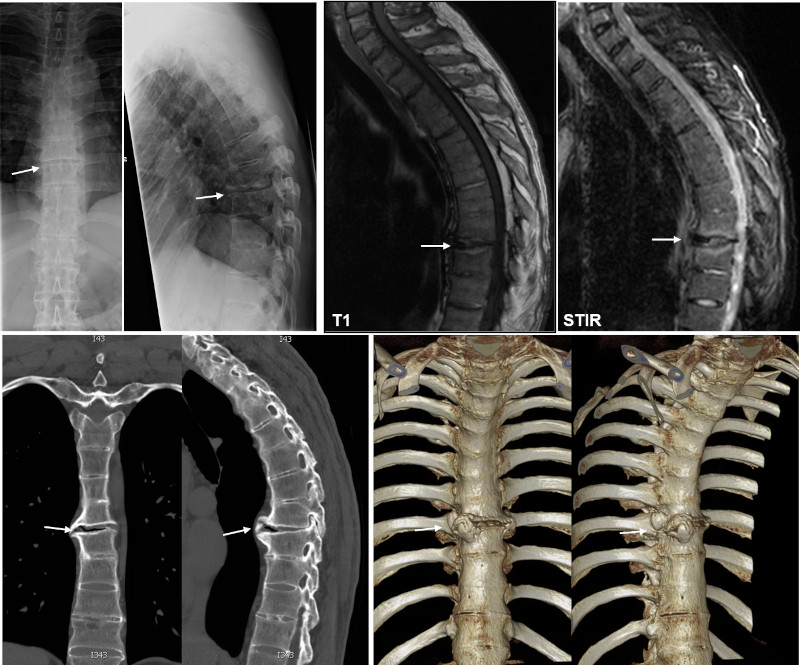
·      Arthritis of costovertebral joints between the posterior lateral aspect of vertebral bodies and the ribs, usually with changes extending to pedicles and posterior aspect of vertebral bodies

·      Arthritis of costotransverse joints lateral to the vertebral bodies with involvement of the rib and transverse vertebral process

·      Enthesitis of spinal ligaments such as the supraspinal and interspinal ligaments

·      Ankylosis/vertebral fusion - new bone formation within the intervertebral disks occurring in long-standing disease.

 
Early AS changes

Corner lesions, relative early active changes, STIR images of the whole spine with enlarged image at the right side showing signs of disease activity in the form of edema at vertebral corners (arrows).


Costovertebral arthritis, sagittal STIR image of the thoracic spine (to the left) shows edema postero-lateral in several vertebral bodies. Supplementary axial STIR images (to the right) illustrate the anatomical location of edema to the joints between the ribs and the vertebral bodies (costovertebral joints) (arrows). This is a typical site for inflammatory changes in AS.

 
Middle stage - mixture of active and structural changes
Sagittal T1-weighted and STIR image of the whole spine to the left with a lateral sagittal STIR image of the thoracic region to the right, showing dispersed corner edema in addition to costovertebral and apophyseal joint inflammation seen on a lateral STIR image. There is additional squaring of the vertebra, especially at the thoracolumbar junction (asterisks) and fat deposition corresponding to vertebral corners anteriorly as well as posteriorly (arrows).
 
Late stage - manifest structural changes

Sagittal T1-weighted  and STIR image of the whole spine to the left with enlargements of the T1 images to the right showing multiple corners with fat deposition in addition to rather general squaring of the vertebrae, especially at the thoracolumbar junction due to osseous destruction of the vertebral corner, best seen corresponding to the intervertebral space Th11-12 (arrow).

 
Late AS stage with deformity

In late stages of AS with spinal deformity it may be impossible to perform an MRI of the entire spine because the patient cannot be positioned within the rather slim MR gantry. However, the lower part of the spine can usually be visualized and if needed supplemented by CT of the upper part as shown beneath.

MRI in a late stage of AS, sagittal T1 and STIR image with supplementary CT of the upper part of the thoracic region showing deformity encompassing reduced lumbar lordosis and increased thoracic kyphosis. There is fusion of L5 and S1 with fat signal across the intervertebral space and anterior vertebral fusion at Th 8-Th11. The supplementary CT of the thoracic region clearly visualizes the vertebral squaring and the universal slim bridging syndesmophytes.

 
Pseudoarthrosis

In late stages of AS with spinal ankylosis pseudoarthrosis may be seen corresponding to intervertebral spaces not ankylosed and thereby being the site of excessive mechanical stress. It can be difficult to evaluate such changes on radiographs and by MRI, but CT will clearly visualize the cleavage and secondary new bone formation as illustrated beneath.

Radiographs, MRI and CT in an AS patient with ankylosed spine except a thoracic intervertebral space without bridging syndesmophytes on the radiographs, but an anteriorly open intervertebral space with subchondral BME on the STIR image (arrows). The CT images, 2D coronal and sagittal reconstructions in addition to 3D reconstruction, clearly show a cleavage with surrounding new bone formation.

 
Grading methods

Grading of inflammatory spinal changes is used to monitor the disease, especially in scientific studies. It is today usually performed in accordance with the SPARCC or Berlin scoring methods as described in the section about MRI (Grading spinal changes by MRI).

 
Visitors since May 1st., 2008:  1765695