The volume of CT data can be reconstructed in multiple planes permitting multiplanar joint assessment. This is especially advantageous for evaluation of the anatomically complex SIJ, often presenting with shadows from overlaying bowel content by radiography. The sensitivity of CT for detecting SIJ changes is therefore higher than by the two-dimensional radiography.
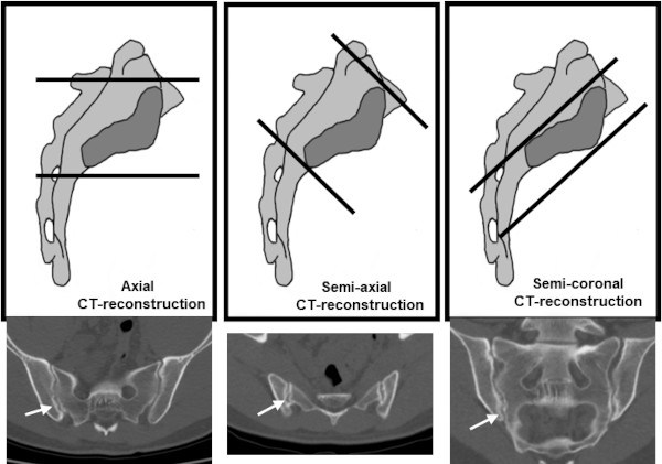
Semi-coronal and semi-axial reconstructions are often used for the interpretation, corresponding to the scan plane used by MRI because the cartilaginous SIJ facets are C-shaped and placed obliquely thereby demanding oblique reconstruction for full visualization of the cartilaginous joint compartment. The original axial CT slices or semi-axial reconstructions are needed for adequate assessment of the ligamentous joint compartment posteriorly.
Original axial CT scan plane and semi-coronal and semi-axial CT reconstructions in relation to the form and orientation of the cartilaginous joint facet. Erosive changes and slight sclerosis (arrows) are seen by all three slice orientations. |