|
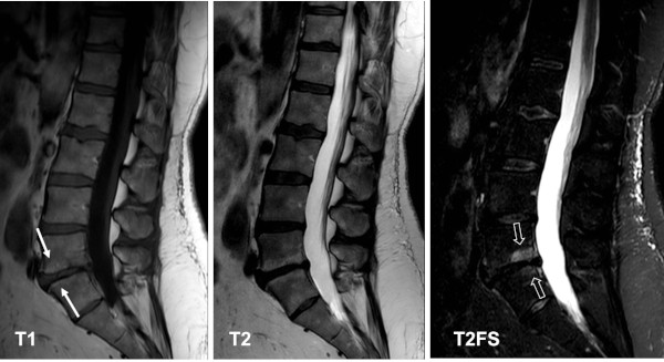
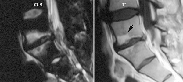
Degenerative spinal changes are frequent causes of back pain. The findings by MRI can vary considerably and there can be subchondral edematous changes as well as fat deposition in accordance with the description of Modic et. al., doi:10.1148/radiology.166.1.3336678.
As part of disk degeneration there can be three different types of endplate changes
Modic type I: represents bone marrow edema with low signal on T1 and high signal on T2 and STIR images.
Modic type II: represents a stage where the changes have converted to fat deposition in the bone marrow with high signal on T1, iso-high signal on T2 and low signal on STIR/T2FS images.
Modic type III: represents subchondral osseous sclerosis with low signal on T1, T2 as well as STIR.
There can be a mixture of the three types, but a common feature for all types is a frequent location along the vertebral plate, often involving the entire subchondral area in contrast to axSpA changes which typically involve the vertebral corners. However, degenerative changes can sometimes predominate at vertebral corners and can then be difficult to differentiate from SpA changes. Besides, patients with axSpA can have concomitant degenerative changes.
Examples of Modic changes are shown below.
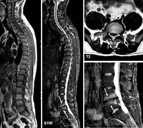
MRI of the entire spine, sagittal T1 and STIR image, in a young patient with inflammatory back pain indicating possible axSpA. There is reduced height and water content corresponding to a lumbar disk with concomitant pronounced subchondral edema encompassing the entire subchondral areas, consistent with Modic type 1 changes (arrows). There is in addition prolapse of the disk, best visualized on the axial T2-weighted slice (open arrows) seen to the right accompanied by a sagittal image showing the image location. |