Dansk  Webmaster  Search   


Technique at radiography
Radiography - sacroiliitis
Radiography - spinal changes
Spinal changes by radiography

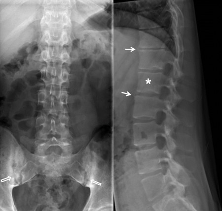
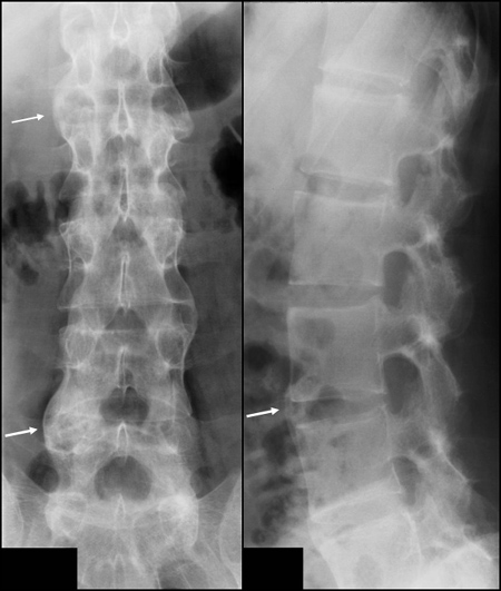
Conventional radiography is a well-established method for detecting structural spinal changes such as new bone formation between the vertebral bodies and vertebral fusion. The slim syndesmophytes characteristic of AS changes are detectable by radiography, but rarely seen by MRI unless being edematous or containing fat deposition.


For the diagnosis of spinal changes a frontal and lateral radiograph (two perpendicular planes) are needed. However, despite using two planes the thoracic spine can be difficult to evaluate on radiographs due to overlaying structures.


Assessment of the severity of spinal abnormalities (grading) in axSpA is usually performed in consistence with the internationally recommended SASS or modified SASS (mSASSS) method https://doi.org/10.1093/rheumatology/key128.

 
Grading methods

SASSS = Stoke Ankylosing Spondylitis Spine Score:


Anterior and posterior changes in the lumbar spine (Th. 12 - S1) at lateral view.

  • Grade 0: Normal findings.
  • Grade 1: Erosion and/or sclerosis and/or squaring.
  • Grade 2: Syndesmophytes (non-bridging).
  • Grade 3: Ankylosis (total bone bridging between upper and lower vertebral edges.

Modified SASSS (mSASSS)

Only anterior changes in both the cervical and lumbar spine at lateral view. Is recommended by OMERACT.

 
Early radiographic changes

Frontal and lateral radiograph of the lumbar spine showing non-bridging syndesmophytes (white arrows) and slight sclerosis at the vertebral corners in addition to slight squaring of the vertebrae, especially the first lumbar vertebra (asterisk). There are bilateral sakroiliit changes (open arrows).

 
Manifest radiographic changes

Frontal and lateral radiograph of the lumbar spine showing bridging syndesmophytes (arrows) between several vertebral bodies.

 
Ankylosis

Frontal and lateral radiograph of the lumbar spine showing fusion of several vertebral bodies (arrow) in addition to fusion of the apophyseal joints and ossification of the interspinous ligaments posteriorly.

 
Visitors since May 1st., 2008:  1766376