|
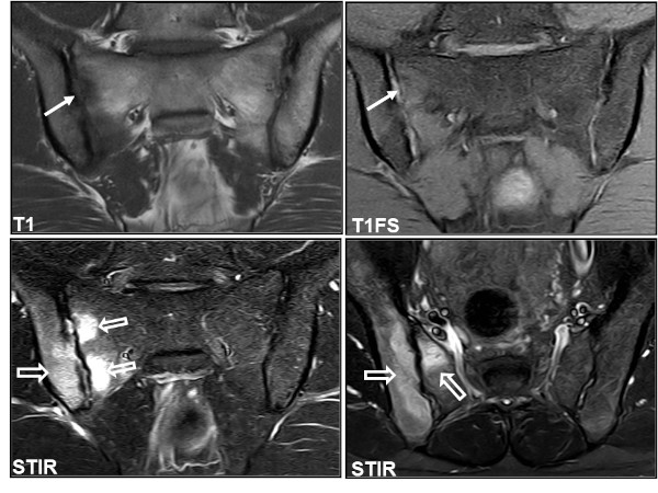
In later stages, MR can provide valuable information about disease activity corresponding to the sacroiliac joints as well as the spine. MRI is therefore also appropriate for estimating the disease activity and can be used to monitor the disease with regard to indication for treatment and for assessing the effect of therapy.
MRI is based on a rather complicated technique. The images can be obtained in numerous ways related to different MR sequences and it is possible to obtain images in arbitrary scan planes. It is important to choose the most appropriate sequences and slice orientation for each specific diagnostic purpose.
During image interpretation, the radiologist must be familiar with normal MRI findings and the appearance of diseases that can simulate sacroiliitis or spinal changes as part of axSpA (differential diagnoses). |