Dansk  Webmaster  Search   


Radiography
CT-scanning
MR-scanning
MRI - technique
MRI-SIJ sequences/planes
MRI - sacroiliitis
Grading sacroiliitis
MRI-Spine sequences/planes
MRI - spinal changes
Grading spinal changes
Spinal changes by MRI

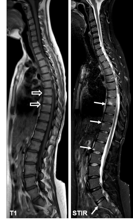
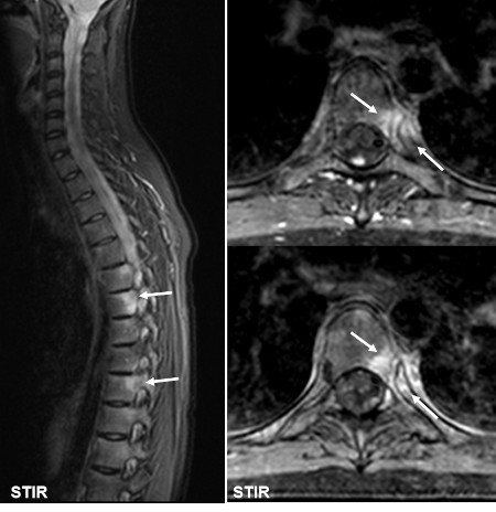
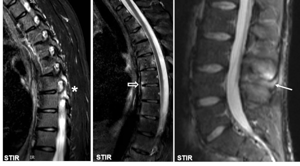
MRI is a sensitive method for detecting active inflammatory changes in the spine presenting as edema in the bone marrow and/or joints. In early stages there is often inflammation corresponding to vertebral corners and there may be signs of inflammation at the costovertebral joints. Later on there may also be active inflammatory changes at the costotransverse and apophyseal joints as well as at the paravertebral ligamentous structures.


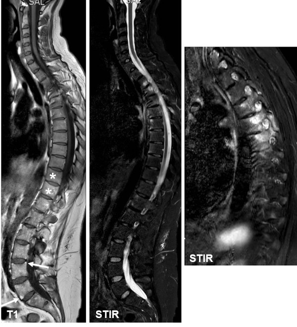
The inflammation at vertebral corner often results in osseous destruction leading to squaring of the vertebral bodies, fat depositions and/or ankylosis across the intervertebral space. New bone formation in the form of syndesmophytes or parasyndesmophytes are, however, often difficult to detect by MRI unless being edematous or containing fat deposition.

Disease activity

Corner lesions, relative early changes, sagittal T1 and STIR image of the whole spine showing signs of disease activity in the form of edema at vertebral corners (arrows) in addition to small fat depositions (open arrows) corresponding to areas with previous active inflammation.


Costovertebral arthritis, sagittal STIR image of the thoracic spine (to the left) shows edema postero-lateral in several vertebral bodies. Supplementary axial STIR images (to the right) illustrate the anatomical location of edema to the joints between the ribs and the vertebral bodies (costovertebral joints) (arrows). This is a typical localization for bone marrow inflammation in ankylosing spondylitis.


Other signs of activity. The upper images show edematous changes corresponding to apophyseal joints (asterisks), syndesmophytes (open arrow) and interspinous ligaments (arrow).

The lower image shows signs of active costotransverse arthritis. Four consecutive sagittal images with the most lateral to the right, visualize edema in the ribs (arrow) with concomitant pronounced soft tissue edema (open arrows).

 
Mixture of active and structural changes

Sagittal T1-weighted and STIR image of the whole spine to the left with a lateral sagittal STIR image of the thoracic region to the right, showing dispersed corner edema in addition to costovertebral and apophyseal joint inflammation seen on a lateral STIR image. There is additional squaring of the vertebra, especially at the thoracolumbar junction (asterisks) and fat deposition corresponding to vertebral corners anteriorly as well as posteriorly (arrow) as a sign of structural changes.

 
Manifest structural changes

Sagittal T1-weighted and STIR image of the whole spine to the left with enlargements of the T1 image to the right showing multiple corners with fat deposition in addition to rather general squaring of the vertebrae, especially at the thoracolumbar junction due to osseous destruction of the vertebral corner, best seen corresponding to the intervertebral space Th11-12 (arrow).

 
Visitors since May 1st., 2008:  1767540